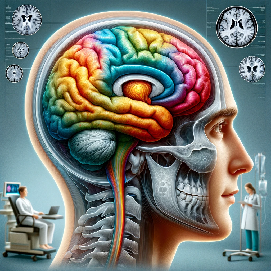
Functional dizziness arises from a complex interaction between the brain’s sensory processing systems, cognitive control networks, and emotional regulation circuits rather than from a single structural lesion or peripheral vestibular disorder. The brain continuously integrates signals from the vestibular organs in the inner ear, visual input from the eyes, and proprioceptive information from muscles and joints to construct a stable sense of orientation in space. In functional dizziness, this multisensory integration process becomes dysregulated, leading to a mismatch between expected and perceived sensory information. The result is a persistent sensation of unsteadiness, lightheadedness, or motion that cannot be fully explained by standard structural or neurological findings.
A central mechanism involves an over-reliance on visual cues for maintaining balance, sometimes referred to as visual dependence. Under normal circumstances, the brain flexibly weights sensory inputs depending on the context, giving more importance to vestibular and proprioceptive information when visual cues are unreliable. In functional dizziness, this adaptive weighting shifts maladaptively toward vision, so that complex or moving visual environments are perceived as excessively destabilizing. This heightened sensitivity to visual motion can intensify symptoms in environments such as supermarkets, busy streets, or scrolling screens, even when objective vestibular function is normal or minimally impaired.
Another key pathophysiological element is the persisting activation of threat and arousal systems within the brain. Following a precipitating event—such as an episode of acute vertigo, a concussion, a panic attack, or a period of intense stress—the brain may continue to interpret normal bodily sensations and minor instabilities as signals of potential danger. This “threat bias” is mediated through limbic structures, including the amygdala, and their connections to vestibular and autonomic centers in the brainstem. The resulting hypervigilance to bodily sensations of dizziness reinforces attention to subtle fluctuations in balance and amplifies the subjective severity of symptoms.
Cortical networks that support spatial orientation and self-motion perception also play a central role. Functional imaging studies suggest abnormalities in connectivity between temporo-parietal regions, which process vestibular and visual motion signals, and frontal areas involved in prediction, attention, and cognitive control. When predictive models in these networks become rigid or inaccurate, the brain may repeatedly generate the expectation of instability or movement even when sensory input indicates relative stability. This prediction error can manifest as a continual feeling of sway, rocking, or internal motion in the absence of actual displacement.
Altered bodily self-awareness, sometimes conceptualized through interoception, further contributes to symptom generation. Individuals with functional dizziness often develop heightened awareness of autonomic responses such as heart rate fluctuations, breathing patterns, and muscle tension. These sensations, normally filtered out as background noise, are interpreted as evidence of imbalance or impending collapse. The dorsal anterior cingulate cortex and insula, regions that integrate interoceptive and emotional information, are thought to be involved in this distorted monitoring of internal bodily states.
Motor control and postural strategies can also become maladaptive. In an effort to prevent falls or manage unpredictability, affected individuals frequently adopt stiffened, guarded postures and reduce natural sway. While this strategy may feel safer, it can paradoxically destabilize balance by limiting the body’s normal automatic adjustments. Over time, these altered movement patterns are reinforced through habit learning within basal ganglia and cerebellar circuits, so that inefficient postural control persists even when the original trigger has resolved.
An important aspect of the pathophysiology involves the transition from an acute to a chronic state. An initial peripheral vestibular insult, orthostatic event, or panic episode may generate legitimate short-term dizziness and imbalance. If during this period the person becomes highly fearful of recurrence, avoids movement, and continually monitors for symptoms, the brain gradually shifts into a state where dizziness is maintained predominantly by central mechanisms. The peripheral insult may improve or resolve, but the central prediction and attention systems continue to generate or amplify sensations of instability, marking the evolution into a functional condition.
Psychological factors are not mere epiphenomena but integral components of the disorder’s physiology. High trait anxiety, intolerance of uncertainty, and catastrophic interpretations of bodily sensations all shape how sensory information is processed. Worry about falling, fainting, or having a serious undiagnosed disease can keep the sympathetic nervous system chronically activated, increasing muscle tension and autonomic reactivity. This, in turn, heightens the perception of bodily movement and lightheadedness, producing a self-perpetuating loop in which anxiety and dizziness continually reinforce each other.
Attentional mechanisms further sustain symptoms by directing disproportionate focus to minor normal fluctuations in stance, vision, and bodily sensations. When attention is narrowly focused on internal cues, external reference points that normally stabilize posture may be disregarded. Neurocognitive models propose that top-down attention from frontal cortical areas biases processing toward internal threat-related signals, while bottom-up sensory inputs that would normally reassure the person of stability are down-weighted. Over time, this skewed attention becomes habitual and automatic.
Neuroplastic changes in central vestibular and visual networks may consolidate these maladaptive patterns. Repeated episodes of visually triggered dizziness and avoidance of movement can strengthen synaptic pathways that associate visual motion with threat and discomfort. Conversely, pathways involved in flexible sensory reweighting, habituation to motion, and confident motor responses may weaken through disuse. This plasticity-based model explains why symptoms can persist for months or years even in the absence of progressive structural pathology, and why systematic rehabilitation can, in many cases, reverse the process.
Autonomic dysregulation is another common dimension of the pathophysiology. Many individuals with functional dizziness display exaggerated heart rate responses, variable blood pressure regulation, or a sensitive vestibulo-autonomic reflex. These autonomic shifts may produce sensations of warmth, palpitations, breathlessness, or near-faintness that the person interprets as evidence of serious imbalance or cerebral hypoperfusion. The interplay between vestibular nuclei and autonomic centers, including the parabrachial nucleus and hypothalamus, provides a biological substrate for the close link between dizziness, nausea, and anxiety symptoms.
Cognitive biases related to expectation and memory help maintain the disorder. After repeated distressing experiences of dizziness in certain environments, the brain forms strong associative memories linking those contexts with danger. Merely anticipating entering a supermarket, standing in a crowded line, or using an escalator can then trigger autonomic arousal and heightened monitoring for signs of instability, which the person experiences as genuine exacerbations of dizziness. This expectancy effect can occur even if objective postural control tests show adequate stability.
Importantly, the pathophysiological mechanisms do not imply that symptoms are imagined or under voluntary control. Instead, they reflect genuine alterations in how the nervous system processes and interprets sensory information, shaped by prior experiences, beliefs, and emotional states. Functional dizziness occupies an intersection between neurology, psychiatry, and otology, where physiological, cognitive, and affective processes are tightly interwoven. These processes produce real, often disabling symptoms despite normal structural imaging and routine laboratory findings.
Because of this multifactorial nature, the condition is best conceptualized within a biopsychosocial framework. Biological predispositions—such as heightened sensory sensitivity or a history of migraine—interact with stressful life events, personality traits, and learned behaviors around health to shape the way the brain handles balance-related signals. Environmental factors, such as visually complex workplaces or prolonged screen use, may further load the system. The pathophysiology thus emerges from dynamic interactions across levels rather than a single fixed lesion.
This understanding helps explain why interventions that target sensory integration, attention, and fear responses can alter the course of the disorder. Techniques such as graded exposure to motion, retraining of gaze and posture, cognitive restructuring of catastrophic beliefs, and anxiety management act directly on the neural circuits and prediction models that generate functional dizziness. By modifying how the brain weighs and interprets vestibular, visual, and proprioceptive input, these approaches leverage neuroplasticity to recalibrate the internal representation of balance and motion.
Clinical features of visual motion sensitivity
Visual motion sensitivity refers to a disproportionate increase in dizziness, unsteadiness, or disorientation when exposed to moving or visually complex environments. Affected individuals commonly describe feeling “off balance,” “swimmy,” or as if the floor is shifting when they are in places with patterned floors, moving crowds, or rapidly changing visual scenes. Symptoms can range from subtle discomfort and mild fogginess to intense vertigo-like sensations, nausea, and fear of falling. Importantly, these symptoms are typically out of proportion to any measurable vestibular deficit, highlighting the functional nature of the disturbance.
Everyday triggers provide a clear window into the clinical picture. Supermarkets and big-box stores are classic examples; long aisles, repeated patterns of shelves, bright overhead lights, and people or carts moving in multiple directions often provoke immediate disequilibrium. Patients frequently report needing to cling to the shopping cart, walk along the wall, or leave the store prematurely. Busy streets, train stations, and airports are similarly challenging, as simultaneous motion of vehicles, escalators, digital displays, and crowds overwhelms visual processing and amplifies the internal sense of motion.
Modern digital environments are another major source of visual motion sensitivity. Scrolling text, rapidly changing images, video calls, first-person video games, and large multi-monitor workstations can provoke eye strain, dizziness, and a sense of veering or rocking. Some individuals notice that turning their head while looking at a screen or watching fast-paced action scenes is particularly destabilizing. This can lead to avoidance of computer work or online communication, with significant occupational and social consequences.
Spatially constrained or patterned environments also feature prominently in symptom reports. Walking down long corridors, especially those with striped carpets or glossy floors that reflect overhead lighting, can produce an illusion of moving walls or an unstable ground surface. People may feel compelled to look down at their feet to compensate, which paradoxically worsens their awareness of subtle body sway. Standing on moving walkways or escalators is often distressing because the apparent visual motion of the environment does not match the expected sense of bodily movement, intensifying the mismatch that characterizes functional dizziness.
Clinically, visual motion sensitivity in functional dizziness has a characteristic temporal profile. Symptoms are usually persistent at a low to moderate level, with superimposed exacerbations when visual motion demands increase. Unlike episodic vertigo caused by conditions such as benign paroxysmal positional vertigo, attacks here are not restricted to brief positional changes but are sustained as long as the provoking environment is present. After leaving a visually demanding setting, patients often describe a lingering after-effect—continued swaying, rocking, or internal motion—that gradually fades over minutes or hours.
The subjective experience of motion is diverse but follows recognizable patterns. Some people feel as if they are being pulled sideways or pushed forward when looking at moving objects, even though they remain standing still. Others describe a sensation that their eyes cannot keep up with the environment, leading to blurring, difficulty focusing, or “tunnel vision.” Spatial disorientation is common: distances may appear distorted, steps may seem higher or lower than they are, and judging the speed of oncoming pedestrians or vehicles becomes challenging. These distortions can increase the risk of cautious missteps, reinforcing the fear of losing balance.
Visual motion sensitivity seldom occurs in isolation and is often accompanied by a constellation of physical symptoms. Nausea, a “hollow” feeling in the stomach, sweating, palpitations, and shortness of breath can arise during exposure to provocative environments. Some individuals experience tightness in the neck and shoulders, headaches or migraine-like pain, and a sense of mental fatigue or “brain fog.” The overlap with migraine features is clinically relevant, as many patients with functional dizziness and visual dependence also have a personal or family history of migraine, even if they do not meet full criteria for vestibular migraine.
From a behavioral standpoint, one of the most striking features is avoidance. To minimize dizziness, individuals gradually restrict activities that involve busy visual scenes: they shop at off-peak hours or rely heavily on online deliveries, avoid public transportation, decline invitations to crowded events, or choose walking routes that bypass busy intersections. Within the home, they may dim lights, limit screen time, or prefer sitting rather than standing when engaged in tasks requiring visual concentration. This avoidance pattern can be subtle at first but becomes entrenched, feeding into the chronicity of symptoms by limiting opportunities for natural habituation and sensory recalibration.
Another prominent clinical characteristic is the adoption of compensatory postural and gaze strategies. Patients often fix their gaze on a non-moving object, such as the floor or a distant point, to reduce the unsettling impact of peripheral motion. They may walk more slowly, keep their feet wider apart, or stiffen their trunk to feel more secure. While these strategies are understandable, they are frequently maladaptive; excessive stiffness interferes with automatic postural reflexes, and persistent downward gaze deprives the balance system of useful horizon and environmental cues. Over time, these patterns become ingrained and contribute to the ongoing perception of instability.
The emotional and cognitive responses to visual motion sensitivity are integral to its clinical presentation. Many individuals become highly vigilant for early signs of dizziness whenever they anticipate entering a visually complex space. This anticipatory anxiety primes the autonomic nervous system, leading to heightened awareness of bodily sensations and a rapid escalation of symptoms. Patients commonly express catastrophic concerns—worries that they may collapse in public, cause an accident, be misjudged by others, or have an undiagnosed serious neurological disease. These thoughts intensify distress and can make relatively mild sensory disruptions feel overwhelming.
In clinical interviews, patients often struggle to find precise language for their experience, alternating between terms like dizziness, lightheadedness, vertigo, imbalance, and “spacey” or “floaty” feelings. They may report that standard questions about spinning sensations or clear positional triggers do not fully capture what they feel. This mismatch can contribute to a sense of not being believed or understood, particularly if previous medical evaluations have focused solely on structural vestibular pathology. Careful, validating exploration of the specific visual triggers, contextual factors, and day-to-day variability in symptoms is therefore essential to articulating the clinical features accurately.
The impact on daily functioning and quality of life is frequently underestimated. Even when physical examination and routine vestibular assessment appear normal, the cumulative burden of restricting shopping, commuting, socializing, and screen-based work can be profound. Fatigue from constant compensatory efforts, loss of confidence in mobility, and the emotional toll of persistent symptoms can contribute to secondary depression and social withdrawal. Understanding these real-world consequences is crucial for planning effective rehabilitation, setting realistic expectations, and motivating patients to engage in interventions that gradually reintroduce controlled visual motion.
Diagnostic criteria and assessment tools
Establishing a diagnosis in patients with functional dizziness and visual motion sensitivity requires a structured, multi-layered approach that integrates symptom characterization, exclusion of significant structural disease, and the identification of positive functional features. The process begins with a detailed clinical history that carefully explores the onset, temporal pattern, and context of dizziness, as well as specific visual motion triggers. Clinicians ask patients to describe in their own words how they feel in environments such as supermarkets, crowded streets, or when using digital screens. The goal is to capture the persistent, fluctuating nature of symptoms, the clear exacerbation in complex visual settings, and the lack of a purely episodic, positional pattern typical of conditions like benign paroxysmal positional vertigo.
Standardized diagnostic criteria have been developed to bring consistency to the diagnosis of functional dizziness, particularly in the form of persistent postural-perceptual dizziness (PPPD), which is the most commonly used framework. According to these criteria, patients experience non-spinning dizziness, unsteadiness, or a sense of internal motion on most days for at least three months. Symptoms are typically aggravated by upright posture, active or passive motion of the body, and exposure to visually complex or moving environments. In addition, the onset frequently follows an identifiable precipitating event, such as an acute vestibular disorder, panic attack, concussion, or significant medical or psychological stressor. Crucially, the symptoms must not be better explained by another primary neurological or vestibular condition, although mild residual deficits from an earlier insult may coexist.
Positive features that support a functional diagnosis extend beyond duration and triggers. The presence of marked visual dependence, with disproportionate distress in situations that involve rich visual motion but relatively preserved function in simple, low-stimulation environments, is one such feature. Another is the mismatch between subjective disability and relatively normal findings on objective tests of balance and ocular motor function. Patients often describe intense swaying or veering sensations despite being able to maintain stance or gait with minimal measurable instability on examination. This dissociation between symptoms and signs, in the absence of inconsistencies suggestive of malingering, is a hallmark of functional dizziness.
A thorough physical and neurological examination remains essential, both to identify comorbid conditions and to reassure patients that dangerous pathology has been appropriately considered. The assessment typically includes bedside tests of vestibular and ocular motor function, such as evaluation of spontaneous and gaze-evoked nystagmus, smooth pursuit, saccades, and the vestibulo-ocular reflex using head impulse testing. Positional maneuvers, including the Dix–Hallpike and supine roll tests, help rule out benign paroxysmal positional vertigo. Screening for cerebellar dysfunction with finger-to-nose, heel-to-shin testing, and tandem gait is important to exclude degenerative or structural disorders. Most individuals with functional dizziness show either normal or only subtly abnormal findings, which can be contextualized for them as evidence of intact structural systems that are being misregulated rather than damaged.
Instrumented vestibular and balance testing may be employed to refine the diagnostic picture and identify coexisting vestibular pathology. Videonystagmography, caloric irrigation, video head impulse testing, rotational chair testing, and vestibular-evoked myogenic potentials can all be used to assess semicircular canal and otolith function. Computerized dynamic posturography is particularly informative in this population, as it quantifies how individuals use visual, vestibular, and proprioceptive cues to maintain balance. In functional dizziness and visual motion sensitivity, posturography often reveals excessive reliance on visual input, with disproportionately poor performance in visually challenging conditions despite relatively preserved stability when vision is removed or altered. These patterns support the concept of visual dependence and maladaptive sensory reweighting rather than structural vestibular failure.
Symptom rating scales and structured questionnaires are valuable tools for quantifying severity, tracking change over time, and identifying co-occurring anxiety or depressive symptoms. The Dizziness Handicap Inventory (DHI) is widely used to assess the perceived impact of dizziness on physical, emotional, and functional domains. High DHI scores in the context of relatively normal objective vestibular test results can reinforce the impression of a functional disorder with significant subjective distress. The Visual Vertigo Analogue Scale, or similar visual motion sensitivity scales, specifically ask patients to rate discomfort in various visually rich scenarios, such as walking through a supermarket, watching moving traffic, or using an escalator. Elevated scores on these instruments corroborate clinical impressions of visual motion sensitivity.
Additional tools include the Situational Characteristics Questionnaire, which systematically catalogs dizziness provocation across different environmental and postural situations, and the Niigata PPPD Questionnaire or other PPPD-specific checklists that align patient symptoms with established diagnostic criteria. For screening broader psychological contributors, brief validated instruments such as the Generalized Anxiety Disorder 7-item scale, Patient Health Questionnaire-9, and measures of health anxiety or catastrophizing can be administered. Elevated anxiety and depressive symptoms do not negate the diagnosis of functional dizziness; instead, they help frame it within a biopsychosocial context and inform subsequent treatment planning.
Visual and oculomotor assessment extends beyond standard neuro-otologic examination in many cases. Some clinicians use dynamic visual acuity tests and gaze-stability assessments during head movement to evaluate functional tolerance of visual motion. Although these may not show frank deficits, patients frequently report subjective discomfort or disorientation when reading or tracking objects while moving their head. Simple in-office tasks, such as asking a patient to stand and turn their head while focusing on a fixed target or to walk while turning their head side to side, can elicit characteristic symptoms of imbalance and visual overload, even if they remain physically stable. Observing the patient’s strategy—such as stiffening the neck, narrowing the visual field, or sharply reducing head movement—provides additional insight into maladaptive compensations.
Neuroimaging is often performed at least once to exclude structural lesions, particularly when symptoms are atypical, there are focal neurological signs, or the patient has significant vascular risk factors. Brain MRI is usually normal or shows only incidental age-related changes in functional dizziness. Communicating the difference between “no findings” and “normal for health and age” is vital, as many patients fear that missed pathology underlies their symptoms. When structural imaging is unremarkable and vestibular tests show minimal or no significant deficits, the clinician can positively frame the diagnosis as one of altered sensory processing and prediction rather than undetected damage, which is a key step in patient education and engagement in rehabilitation.
Clinical interviews should explicitly probe for the temporal relationship between dizziness and potential triggers or stressors. Questions about recent infections, head injuries, episodes of acute vertigo, panic attacks, medical procedures, or major life events often reveal a clear precipitating event that aligns with PPPD criteria. It is equally important to explore the course of symptoms, including whether they improved then plateaued, whether new avoidance behaviors emerged, and how anticipatory anxiety shapes day-to-day decisions. Mapping these trajectories helps differentiate functional dizziness from progressive neurodegenerative or structural conditions, which typically show a different temporal evolution.
Another core component of the diagnostic process is evaluating functional impact. Clinicians should ask specifically about shopping, driving, public transportation, screen use, work performance, and social participation. Patients are encouraged to describe activities they have stopped, modified, or endure with significant distress because of visual motion sensitivity. This functional history not only supports the diagnosis but also sets a baseline for outcome measurement during vestibular therapy and psychological interventions. Furthermore, it clarifies priorities for individualized rehabilitation goals, such as returning to work, tolerating supermarket visits, or resuming public transit use.
Because functional dizziness exists at the interface of neurology, otology, and psychiatry, collaborative assessment across specialties can be highly beneficial. A neurologist or otologist may lead the structural and vestibular workup, while a psychologist or psychiatrist evaluates health beliefs, coping strategies, and coexisting mood or anxiety disorders. Physical therapists with expertise in vestibular rehabilitation can perform detailed gait and balance assessments, including analysis of posture, head movement, and environmental interaction. When findings from different disciplines converge on a pattern of persistent, visually aggravated dizziness with largely preserved structural function, confidence in the functional diagnosis increases and allows the care team to present a unified explanation to the patient.
Patient education is woven throughout the assessment process and serves both diagnostic and therapeutic purposes. Clinicians can gauge understanding and beliefs by asking what the patient thinks is causing the dizziness, what they fear most, and how they imagine their balance system works. Misconceptions—such as the belief that any dizziness implies brain damage or impending stroke—are common. Correcting these in real time using simple explanations about sensory integration, visual dependence, and functional nervous system changes helps shift the narrative away from undetected structural disease and toward a model that is compatible with recovery. A patient who can articulate, in their own words, that their symptoms reflect “an over-sensitive balance system that relies too much on vision” is more likely to engage constructively with rehabilitation.
In practice, the diagnosis is ultimately a synthesis of all these elements: chronic, non-spinning dizziness and unsteadiness; exacerbation by upright posture, motion, and complex visual environments; evidence of visual motion sensitivity and maladaptive postural strategies; a history of precipitating events or psychological stressors; relatively normal or only mildly abnormal vestibular and neurological findings; and significant functional impairment disproportionate to structural pathology. When these features are present, clinicians can make a confident diagnosis of functional dizziness, often within the PPPD framework, and proceed to discuss evidence-based treatment options that emphasize graded exposure, vestibular rehabilitation, and cognitive-behavioral strategies rather than further rounds of purely diagnostic testing.
Evidence-based treatment approaches
Management of functional dizziness and visual motion sensitivity is most effective when it combines education, vestibular therapy, graded exposure to visual motion, and psychological interventions that address maladaptive beliefs and anxiety. Treatment is not aimed at “finding and fixing” a damaged structure but at recalibrating how the brain processes sensory information, reallocates attention, and responds to bodily signals of threat. Because multiple systems are involved, an interdisciplinary approach that may include otolaryngologists, neurologists, vestibular physical therapists, psychologists, and sometimes occupational therapists generally yields the best results.
Clear, structured education is a cornerstone of care and begins with an explanation that symptoms are real but arise from functional changes in sensory integration rather than irreversible damage. Clinicians can describe how the brain has become over-reliant on visual input and hypervigilant to internal sensations, leading to exaggerated dizziness and imbalance in complex environments. Using familiar examples—such as feeling off balance in a supermarket or when scrolling on a phone—helps patients connect the explanation to their own experience. The educational message emphasizes that the nervous system remains capable of change, that recovery is typically gradual, and that active participation in rehabilitation is crucial. This reframing alone can reduce catastrophic misinterpretations and lower baseline anxiety, making subsequent interventions more effective.
Vestibular rehabilitation is the primary physical treatment modality and is strongly supported by clinical evidence for PPPD and related functional dizziness conditions. Individualized vestibular therapy programs are designed and supervised by trained vestibular physical therapists, with exercises targeting gaze stabilization, postural control, and sensory reweighting. Gaze-stability training might include focusing on a target while the head is moved horizontally or vertically, first in sitting, then in standing, and eventually during walking. Postural exercises progress from stable to unstable surfaces, from wide to narrow stance, and from eyes open to eyes closed. The goal is to reduce visual dependence and strengthen the use of vestibular and proprioceptive cues for balance.
A critical component of vestibular therapy for visual motion sensitivity is systematic habituation to visually complex stimuli. Rather than avoiding provoking environments, therapy introduces them in a graded, controlled manner. Early stages may involve watching simple patterns on a small screen or slowly scrolling text while seated. Over time, patients are guided to tolerate more challenging visual motion, such as videos of crowds or moving traffic, walking through visually busy corridors, and eventually visiting supermarkets or shopping centers. Exposures are kept below a level that triggers overwhelming distress, and patients are taught to remain in each situation long enough for symptoms to peak and then begin to diminish, which promotes habituation and recalibration of sensory weighting.
Task-specific functional training bridges the gap between clinic-based exercises and real-world participation. Therapists collaborate with patients to identify high-priority activities, such as grocery shopping, using public transit, working at a computer, or attending social events. These tasks are then broken into manageable steps. For example, a supermarket plan might start with standing near the entrance for a short time, progress to walking one aisle with a cart, and then expand to a full shopping trip during busier hours. The therapist helps the patient monitor symptom intensity, use adaptive breathing and grounding strategies, and recognize gradual improvements across sessions. This approach counters avoidance, builds confidence, and directly targets the contexts in which functional dizziness is most disabling.
Evidence-based psychological treatments, particularly cognitive-behavioral therapy (CBT), play a central role in addressing the cognitive and emotional processes that maintain symptoms. CBT protocols for functional dizziness focus on identifying catastrophic interpretations of dizziness (for example, “If I feel unsteady, I will collapse or have a stroke”), challenging these thoughts using evidence from past experiences and medical assessment, and replacing them with more balanced appraisals (“These sensations are a sign that my balance system is over-alert, not that I am in immediate danger”). Therapists work with patients to map the vicious cycle in which dizziness leads to fear, hypervigilance, muscle tension, and further dizziness, and then develop strategies to interrupt this loop.
Anxiety-management techniques are integrated into CBT and rehabilitation. Patients are taught controlled breathing, progressive muscle relaxation, and mindfulness-based attention training to reduce physiological arousal when entering visually challenging situations. Mindfulness exercises emphasize noticing sensations of motion or imbalance without immediately reacting with fear or escape behavior, which can gradually weaken the learned association between dizziness and danger. Some programs use acceptance and commitment therapy (ACT) principles to help patients engage in valued activities despite residual symptoms, reinforcing the message that they can live well while the nervous system continues to recalibrate.
Graded exposure, a shared feature of both vestibular rehabilitation and CBT, is particularly important in treating visual motion sensitivity. In addition to physical exposures to busy environments, therapists may use imaginal and virtual exposures. For instance, patients might visualize walking through a crowded train station while practicing calm breathing, or use virtual reality headsets to experience controlled visual motion in a safe clinic setting. Research indicates that such exposures reduce avoidance, diminish anxiety responses, and improve functional tolerance of complex visual scenes, supporting their inclusion in comprehensive treatment plans.
Pharmacologic interventions can be helpful as adjuncts when anxiety, depression, or migraine significantly complicate recovery, though they are rarely sufficient as stand-alone treatments. Selective serotonin reuptake inhibitors (SSRIs) and serotonin–norepinephrine reuptake inhibitors (SNRIs) have shown benefit in PPPD, likely by dampening central hypervigilance and threat responses and improving mood. When used, medications are introduced at low doses and titrated slowly to minimize side effects that might mimic or worsen dizziness. Benzodiazepines, while sometimes prescribed for acute vertigo, are generally avoided in persistent functional dizziness because they can impair central compensation, promote dependence, and interfere with learning during rehabilitation.
Because many individuals with functional dizziness have comorbid migraine, especially vestibular migraine, migraine-directed therapies may also form part of the treatment strategy. Lifestyle measures such as regular sleep, hydration, and meal timing, along with prophylactic medications when indicated, can reduce overall sensory sensitivity and headache burden. As migraine control improves, tolerance of visual motion often increases, allowing more intensive participation in vestibular and psychological rehabilitation. Coordination between the treating neurologist and vestibular therapist ensures that adjustments in migraine management are aligned with ongoing balance-focused interventions.
Occupational therapy can provide targeted support for patients whose work or daily routines are heavily screen-based or visually demanding. Occupational therapists may recommend modifications such as adjusting monitor size and distance, altering font size and contrast, using screen filters, implementing structured breaks, and redesigning task workflows to minimize rapid visual shifts. They also help patients rehearse ergonomic postures and gaze strategies that facilitate balance while maintaining productivity. These practical adjustments can reduce symptom provocation during essential activities, preventing secondary disability and job loss.
Self-management strategies are emphasized throughout treatment to reinforce gains and promote long-term resilience. Patients are encouraged to maintain regular physical activity within tolerable limits, as deconditioning can worsen fatigue and perceived imbalance. They may keep brief activity and symptom logs to identify progress and patterns, while avoiding excessive monitoring that could increase preoccupation with dizziness. Sleep hygiene, moderate caffeine and alcohol use, and stress-management practices support nervous system stability. Clinicians often provide written home programs summarizing key exercises, exposure steps, and cognitive strategies, empowering patients to continue rehabilitation outside of formal sessions.
Collaborative goal setting ensures that treatment remains meaningful and patient-centered. Early in the process, clinicians ask patients to identify specific activities they want to regain—such as shopping independently, attending a family gathering, or driving on a busy highway. These goals are translated into concrete, measurable steps that can be tracked over time. Regular review of progress helps sustain motivation, validates effort, and allows timely adjustments to the treatment plan when barriers arise. By linking exercises and exposure tasks to personally important outcomes, rehabilitation becomes more engaging and less abstract.
Family education and involvement can further enhance treatment effectiveness. Partners or relatives often witness the patient’s difficulties in busy environments and may unintentionally reinforce avoidance by taking over tasks or overprotecting the individual. Clinicians can explain the functional nature of dizziness, the rationale for gradual exposure, and the importance of encouraging—not forcing—participation in challenging activities. Family members learn how to provide calm, supportive presence during exposures, help track small improvements, and avoid alarmist reactions to transient symptom flares. This supportive context can reduce interpersonal stress and create a home environment that aligns with therapeutic goals.
Across modalities, treatment success depends on consistency and pacing. Interventions are most beneficial when practiced regularly, ideally daily or several times per week, with modest, incremental increases in difficulty. Pushing too aggressively can provoke discouraging symptom spikes, while progressing too slowly may allow avoidance patterns to persist. Clinicians coach patients to expect temporary symptom increases during new tasks and to interpret these not as signs of harm but as evidence that the balance system is being challenged in a way that promotes adaptation. Over weeks to months, many patients report that episodes of dizziness become shorter, less intense, and less frightening, and that they can remain active in situations that were previously intolerable.
Long-term outcomes and prognosis
Long-term outcomes in functional dizziness and visual motion sensitivity are generally favorable when the condition is correctly identified and managed with a comprehensive, individualized plan. Many individuals experience substantial reductions in symptom intensity, improved tolerance of visually complex environments, and a return to regular daily activities. However, progress is often gradual rather than rapid, typically unfolding over months instead of days or weeks. Understanding this time course is crucial for setting realistic expectations and preventing discouragement when dizziness fluctuates during rehabilitation.
Follow-up studies in populations meeting criteria for persistent postural-perceptual dizziness suggest that a significant proportion of patients achieve meaningful functional recovery. Those who engage consistently in vestibular therapy and cognitive-behavioral interventions, and who adhere to graded exposure to provocative visual motion, tend to show the greatest gains. Improvements may first be noticed as shorter recovery times after exposure to busy environments, followed by an increased ability to remain in such environments without needing to escape. Eventually, many people can resume activities like supermarket shopping, public transport use, and screen-based work with only mild, manageable discomfort or no symptoms at all.
Despite this generally encouraging outlook, complete symptom resolution is not universal. A subset of individuals continues to experience intermittent episodes of unsteadiness or visual disorientation, particularly during periods of stress, fatigue, or illness. For these patients, long-term success is often defined not by the absolute disappearance of symptoms but by a shift in how symptoms are interpreted and managed. When dizziness is recognized as a benign signal of a temporarily overactive balance system rather than a marker of serious disease, its impact on functioning diminishes. People learn to implement brief self-directed exercises, pacing strategies, and cognitive techniques that allow them to continue their activities rather than withdrawing.
The durability of treatment gains is closely linked to whether patients integrate new habits into their daily routines. Those who maintain regular physical activity, continue occasional balance and gaze-stability exercises, and periodically practice exposure to challenging environments are more likely to sustain improvements. In contrast, prolonged avoidance of visually rich settings after formal therapy has ended can gradually erode gains and permit old patterns of visual dependence and hypervigilance to reemerge. Clinicians therefore emphasize that rehabilitation is not a one-time event but a set of skills that can be revisited and adjusted across the lifespan as circumstances change.
Psychological factors play a major role in long-term prognosis. High baseline health anxiety, severe generalized anxiety, or untreated depression are associated with more persistent symptoms and greater functional impairment over time. When these conditions are adequately addressed through therapy and, when appropriate, medication, the outlook for functional dizziness improves substantially. Conversely, ongoing catastrophic interpretations of bodily sensations, rigid beliefs about fragility, and repeated reassurance-seeking from emergency services or multiple specialists can perpetuate the disorder. Early, clear explanations of the diagnosis and timely referral to appropriate psychological care help reduce the risk of chronicity.
Comorbidities such as migraine, orthostatic intolerance, and other functional somatic symptoms also influence long-term outcomes. Individuals with well-controlled migraine or autonomic dysfunction typically report more stable trajectories, whereas poorly managed comorbid conditions can provoke symptom flares even after successful initial rehabilitation. In integrated care models, neurologists, cardiologists, and primary care providers collaborate with vestibular and psychological specialists to optimize management of these coexisting issues. When the broader health context is stabilized, the nervous system is better able to consolidate the sensory and cognitive changes achieved during treatment.
Work status and social participation are key indicators of long-term prognosis. Many patients who initially reduce work hours or take medical leave due to visual motion sensitivity can gradually return to employment, especially when workplace accommodations are implemented. Flexible scheduling, modified visual demands, and staged increases in workload allow the balance system to adapt without overwhelming it. Remaining engaged in at least some level of work or structured activity during rehabilitation is associated with better long-term adjustment, whereas prolonged disengagement tends to lower confidence and heighten focus on bodily sensations.
Relapse patterns, when they occur, often follow predictable triggers. Major life stressors, acute illnesses, or abrupt increases in visually demanding tasks—such as starting a new job with intensive screen use—can provoke a temporary resurgence of symptoms. However, individuals who have previously completed vestibular therapy and psychological treatment usually possess tools to manage these setbacks more effectively than before. They may reintroduce specific exercises, temporarily scale back exposure intensity, and apply cognitive restructuring techniques to prevent escalation into a renewed cycle of avoidance and fear.
Long-term follow-up appointments or periodic check-ins with members of the treatment team can reinforce progress and provide an opportunity to fine-tune self-management plans. During these visits, clinicians may repeat targeted elements of the original assessment, such as brief balance tests or symptom questionnaires, to document changes over time. Demonstrating measurable improvement in objective and subjective domains can strengthen patients’ confidence in their own capacity for ongoing adaptation, even if occasional dizziness persists. These sessions also offer a forum for troubleshooting new challenges, such as changes in work demands or aging-related shifts in physical capacity.
Age itself does not preclude favorable outcomes, although the trajectory may differ between younger and older individuals. Younger patients may adapt more quickly to intensive exposure-based programs and regain high levels of activity, but they can also be more distressed by disruptions to work, study, or social life. Older adults may progress more slowly due to comorbid musculoskeletal or cardiovascular conditions, yet they often benefit greatly from tailored, lower-intensity rehabilitation that supports safe mobility and independence. Across age groups, individualized pacing and realistic, function-oriented goals are more predictive of success than any single demographic factor.
The quality of the initial diagnostic interaction exerts a lasting influence on prognosis. When clinicians offer a coherent explanation, validate the reality of symptoms, and outline a clear path forward, patients are more likely to trust the diagnosis and commit to active rehabilitation. In contrast, fragmented or contradictory messages—such as alternating reassurances that “nothing is wrong” and suggestions of serious but unspecified disease—can foster doubt and ongoing medical shopping. A stable, well-communicated conceptualization of functional dizziness as a reversible problem in sensory processing helps anchor treatment efforts and improves long-term engagement.
Socioenvironmental factors also shape long-term outcomes. Individuals with strong social support, stable housing, and access to specialized care tend to fare better than those facing financial strain, unstable living conditions, or limited healthcare resources. When possible, connecting patients with community resources, support groups, or online education materials can help bridge these gaps. Peer support, whether in-person or virtual, offers validation, practical coping ideas, and a sense of shared experience that can reduce isolation and promote perseverance with rehabilitation strategies.
In a broader sense, prognosis improves as understanding of functional neurological and vestibular disorders progresses within the healthcare system. Increased clinician familiarity with diagnostic criteria, assessment tools, and evidence-based interventions means that patients are more likely to receive timely, appropriate care rather than undergoing prolonged, inconclusive investigations. Earlier recognition reduces the period during which maladaptive patterns of avoidance, hypervigilance, and visual dependence can become entrenched, which in turn leads to better long-term trajectories. As interdisciplinary clinics and standardized treatment pathways become more common, the expectation of meaningful improvement—rather than resignation to lifelong disability—can be more confidently communicated to patients.
Although longitudinal data on very long-term outcomes are still developing, existing evidence and clinical experience point to a pattern in which functional dizziness transitions from a dominating, unpredictable problem to a more manageable, background issue or, for many, recedes almost entirely. The central determinants of this transition include early accurate diagnosis, engagement with structured vestibular and psychological rehabilitation, attention to comorbid conditions, and sustained use of self-management skills. When these elements come together, individuals can typically maintain active, fulfilling lives even in the face of occasional residual sensations of imbalance or visual discomfort.