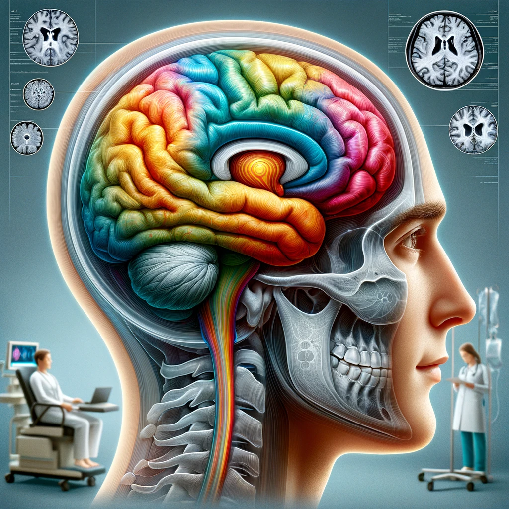
A functional neurological disorder is defined by genuine neurological symptoms that arise from altered functioning of the nervous system rather than from structural damage or a degenerative process. The defining feature is the presence of motor, sensory, or cognitive changes that resemble recognized neurological diseases yet are internally inconsistent or physiologically incongruent when examined carefully. This is not a diagnosis of exclusion; it rests on positive clinical features that indicate a problem in how the brain is operating, not on the absence of abnormalities alone.
Terminology has evolved from the older label conversion disorder to contemporary usage that emphasizes mechanism and clinical phenomenology. Functional neurological disorder (often abbreviated FND) is the umbrella term used in neurology, while psychiatric classification systems may refer to functional neurological symptom disorder. The condition sits at the interface of neurology and psychiatry, an area commonly described as neuropsychiatry, reflecting the interplay between brain networks, cognition, emotion, and behavior in shaping symptoms.
Several principles distinguish FND from other conditions. First, the symptoms are involuntary and not fabricated or feigned; they are experienced as real and distressing. Second, the clinical picture includes rule-in signs such as variability over short time frames, improvement with distraction, or patterns that do not conform to known lesion-based neuroanatomy. Third, a person can have FND alongside another neurological disease, meaning the presence of a structural diagnosis does not exclude a concurrent functional one.
The symptom spectrum is broad. Common presentations include limb weakness or paralysis, tremor and other movement disorders, gait disturbance, functional seizures (also called dissociative or nonepileptic attacks), sensory loss or altered sensation, visual symptoms such as intermittent visual loss, speech and voice changes, and cognitive difficulties like impaired concentration. Symptoms often fluctuate in intensity, may be triggered or worsened by attention and expectation, and can cluster with pain, fatigue, sleep disturbance, and autonomic complaints.
The definition emphasizes mechanisms of dysfunction rather than a requirement for a specific stressor. Although some individuals report precipitating events such as injury, illness, or psychological stress, many do not identify a single cause. Risk and maintaining factors can include prior adverse experiences, heightened symptom vigilance, illness beliefs, physiological arousal, and reinforcement processes, but none are necessary to establish the diagnosis. This approach moves beyond the outdated notion of medically unexplained symptoms by prioritizing observable clinical features that support a functional mechanism.
Language is central to the definition. Functional indicates a problem in how neural networks are operating rather than a structural lesion, and the term is intended to be descriptive and nonjudgmental. Clear communication that the disorder is common, real, and potentially reversible helps align expectations and promotes engagement. Framing the condition as a disorder of brain function provides a platform for collaborative diagnosis and treatment without implying blame or intentional control.
In clinical practice, defining FND involves recognizing characteristic patterns that are incongruent with known disease pathways, identifying positive bedside signs, and mapping the symptoms to a coherent functional formulation. This formulation integrates biological vulnerability, psychological context, and social factors to explain why the symptoms are present and persist, guiding individualized treatment while maintaining a neurological standard of assessment and follow-up.
Because the definition rests on positive evidence of functional mechanisms, it enables earlier diagnosis, reduces unnecessary investigations, and opens a pathway to evidence-based treatment that targets retraining of movement and perception, optimization of attention and expectation, and management of comorbidities. This definition underscores that recovery is feasible and that prognosis is closely linked to recognition, explanation, and tailored intervention.
Mechanisms and pathophysiology
Mechanistic models emphasize dysfunction of distributed brain networks rather than damage to a single node. In a functional neurological disorder, perceptual, motor, and interoceptive systems appear to misweight expectations and sensory evidence, so that top-down predictions gain excessive influence over moment-to-moment experience. The consequence is the emergence of involuntary symptoms that are internally coherent to the brain’s predictive model but incongruent with lesion-based neuroanatomy. This framework helps explain why symptoms can be variable, context-dependent, and influenced by attention and expectation.
Predictive processing accounts propose that the brain constantly generates hypotheses about incoming signals and assigns “precision” (confidence) to predictions and sensory inputs. When precision is abnormally allocated—often amplified by heightened self-monitoring or threat appraisal—predictions can dominate perception and motor output. In the motor system, this may manifest as inhibited or competing motor programs that interfere with intended movement, while in sensory domains it can produce altered sensation or loss of function without structural pathology. The person experiences these effects as real and involuntary because the underlying inference processes occur outside conscious control.
Evidence from functional neuroimaging supports network-level alterations in FND. Studies frequently show increased activity and connectivity within salience and limbic circuits (including amygdala, insula, and anterior cingulate) alongside changes in sensorimotor regions such as the supplementary motor area, premotor cortex, and cerebellum. At the same time, regions involved in monitoring agency and integrating multisensory signals—such as the temporoparietal junction and posterior parietal cortex—may be under-engaged during symptomatic states. Heightened salience signaling can bias attention toward internal bodily signals, while altered parietal integration can degrade the sense that movements are self-generated, contributing to the experience of loss of control.
Abnormalities in arousal and interoception appear integral to pathophysiology. The insula, a hub for integrating visceral signals with cognitive and emotional context, shows altered activation and connectivity, suggesting that internal bodily cues may be interpreted as more threatening or salient. Changes in noradrenergic and hypothalamic-pituitary-adrenal stress systems can increase bodily arousal, amplifying prediction errors that reinforce symptoms. These processes do not require an external stressor at onset, but they can maintain symptom cycles once established.
Learning mechanisms and reinforcement further shape persistence. After an initial precipitant—such as injury, illness, or an episode of acute stress—associations can form between specific movements, sensations, or contexts and perceived danger or failure. Avoidance, safety behaviors, and repeated symptom-focused attention strengthen these associations through classical and operant conditioning. Over time, habits within cortico-striato-thalamo-cortical loops may consolidate maladaptive motor patterns, while cerebellar circuits that ordinarily fine-tune predictions become calibrated to dysfunctional priors.
Physiological studies of motor control illustrate how bedside signs arise from these mechanisms. In functional weakness, for example, apparently impaired voluntary drive can coexist with preserved automatic motor output, as seen when hip extensors activate normally during contralateral hip flexion even if voluntary extension seems weak. Functional tremor often entrains to external rhythms and may change with distraction, consistent with a system in which attention alters the gain on competing oscillatory outputs. In functional jerks, some recordings reveal premovement potentials compatible with an involuntary, preplanned action that escapes conscious agency. Across phenotypes, corticospinal tracts and neuromuscular transmission are typically intact, underscoring that the disturbance lies in coordination, selection, and gating of motor commands.
Sensory manifestations reflect similar top-down influences. Functional sensory loss can show boundaries aligned with attentional representations rather than dermatomes, and visual symptoms may fluctuate with cognitive load or expectation. Altered connectivity between visual or somatosensory cortices and salience networks suggests that predictions about threat or impairment shape how ambiguous sensory input is perceived, generating convincing but reversible deficits.
In functional seizures, network dynamics point to a transient reconfiguration of attention, salience, and motor systems rather than epileptiform hypersynchrony. During episodes, impaired integration between frontal control networks and motor output, coupled with heightened arousal and dissociation, can produce unresponsiveness or bilateral movements without the EEG signatures of epilepsy. Post-event fatigue and cognitive fog may reflect the metabolic and autonomic costs of these network shifts rather than neuronal injury.
Individual vulnerability likely arises from a mix of biological and experiential factors—such as trait anxiety, prior adverse experiences, neurodevelopmental differences, pain, fatigue, or concurrent medical illness—that bias precision weighting, threat appraisal, and attentional capture. However, none of these are necessary or sufficient on their own, and many people with FND report no clear precipitant. The heterogeneity of mechanisms explains why presentations vary and why neuropsychiatry approaches emphasize formulation over a single cause.
Understanding these mechanisms has practical implications for diagnosis and treatment. Positive bedside signs reflect modifiable network behavior, allowing clinicians to demonstrate reversibility during examination. Interventions that redirect attention, recalibrate predictions, and rebuild automaticity—such as specialized physiotherapy, motor retraining, exposure-based strategies, and targeted psychological therapies—map directly onto the involved circuits. Education that frames symptoms as real outputs of a miscalibrated but plastic system can reduce threat signaling, improve engagement, and create conditions for neural relearning.
Clinical features and differential diagnosis
Clinical assessment hinges on recognizing positive signs that point toward a functional neurological disorder rather than relying solely on exclusion of other diseases. Across presentations, internal inconsistency, variability over short time frames, and changes with distraction or focused attention are central clues. Many people demonstrate preserved automatic function despite impaired voluntary control, and symptom expression often shifts with context, task demands, or testing maneuvers that redirect attention or alter expectations.
Functional limb weakness typically shows patterns incongruent with corticospinal lesions. Give-way or collapsing weakness may appear with sustained testing, yet strength improves when the same muscle is recruited automatically. Hoover’s sign—where apparent hip extension weakness improves during contralateral hip flexion against resistance—exemplifies preserved involuntary motor output. The hip abductor sign and improvement of drift with distraction provide additional rule-in evidence. Reflexes, tone, and pyramidal signs are usually normal, and distribution of weakness may not fit a single root, nerve, or cortical territory.
Functional tremor is often the most common movement presentation. Key features include distractibility, sudden onset, variability in frequency and amplitude, and entrainment to the rhythm of a tapping task performed with the contralateral limb. Co-contraction of antagonist muscles and abrupt pauses during cognitive tasks may be observed. Unlike Parkinson disease, rest tremor may vanish during unobserved periods and reappear with attention. Functional myoclonus and jerks frequently show stimulus sensitivity and a preplanned quality on examination, with latency and variability inconsistent with reflex myoclonus. Fixed postures (often termed functional dystonia) may arise suddenly after minor injury and remain immobile; unlike organic dystonia, classic sensory tricks are less reliably helpful.
Gait disturbance in FND ranges from knee buckling with rare true falls to a lurching, variable base with exaggerated sway that improves when attention is diverted. Some individuals exhibit a dragging monoplegic gait with the forefoot in contact with the ground, yet can briefly generate normal step height during dual-tasking. Start-and-stop phenomena, excessive slowness without freezing, and improved balance during complex but engaging tasks (e.g., dancing, marching to a beat) support a functional mechanism.
Sensory symptoms often display nondermatomal or sharply demarcated boundaries that respect attentional maps more than anatomy, such as midline-splitting of facial or body sensation. Vibration sense may seem absent at the ankle yet be perceived at the tibial crest, and light touch can feel painful or diffuse. Visual presentations can include intermittent visual loss, concentric “tubular” fields that remain the same size regardless of test distance, or variable diplopia that does not align with a single cranial nerve distribution. These patterns are compelling when they fluctuate during the same visit or improve with reassurance and guided attention.
Speech and communication features include functional dysphonia with a whispering or hoarse voice that improves during coughing or laughing, intermittent stuttering that disappears with singing, and variable dysarthria that waxes and wanes within minutes. Cognitive complaints commonly involve concentration and working memory lapses that worsen under scrutiny but improve in low-pressure contexts; bedside testing may reveal inconsistent effort with better performance on complex tasks than on simpler ones when attention is reframed.
Functional seizures (also called dissociative or nonepileptic attacks) have characteristic semiology. Episodes often last longer than typical generalized tonic-clonic seizures, show waxing and waning intensity, involve closed eyes with resistance to passive opening, side-to-side head movements, pelvic thrusting, or asynchronous limb activity, and preserve protective responses (e.g., avoiding injury) despite apparent unresponsiveness. Post-event confusion may be brief or absent, and tongue biting, when present, more commonly involves the tip rather than the lateral tongue. A mixed picture can occur, and some individuals with FND also have epileptic seizures, making careful documentation of typical events essential for accurate diagnosis and treatment planning.
Differential diagnosis depends on the presenting system. In suspected stroke or transient ischemic attack, isolated weakness with normal reflexes and tone, rapid fluctuations, and improvement with contralateral activation argue against an acute vascular lesion; nonetheless, new focal deficits or vascular risk factors warrant standard stroke workup. In neuromuscular disorders such as myasthenia gravis or neuropathies, fatigable weakness, objective fatigability on repetitive testing, and consistent distribution patterns are expected; in contrast, FND shows improved performance with distraction and inconsistent fatigability. For movement disorders, Parkinson disease features bradykinesia with decrement and rigidity, whereas functional tremor shows entrainment and distractibility; essential tremor has a relatively fixed frequency with family history and alcohol responsiveness, findings that do not define functional tremor.
In sensory and visual complaints, peripheral neuropathy yields length-dependent loss and reproducible deficits on quantitative testing, while FND presents with shifting borders and mismatch between modalities. Multiple sclerosis or other demyelinating diseases typically show objective signs, MRI lesions, or evoked potential abnormalities; a functional picture lacks these correlates and displays positive inconsistency signs. Vestibular disorders generally produce nystagmus patterns and postural responses that follow physiological rules; in functional dizziness, sway patterns and symptom intensity are highly context dependent and may improve during dual-tasking.
Distinguishing FND from factitious disorder or malingering rests on the clinical hallmarks of involuntariness and positive rule-in signs. Symptoms in FND are experienced as genuine and distressing, fluctuate with attention and expectation, and improve with techniques that restore automaticity; deliberate feigning is not supported by these findings. Coexisting conditions are common, including migraine, pain syndromes, hypermobility, anxiety, depression, posttraumatic stress, and dysautonomia; their presence does not negate a functional mechanism and may shape formulation and management priorities.
Red flags for alternative or concurrent disease include progressive stepwise deterioration without fluctuation, objective upper motor neuron signs, consistent focal deficits that map to neuroanatomy, unexplained weight loss or systemic features, new-onset headaches with red flags, and laboratory or imaging abnormalities that fit the clinical picture. Conversely, rapid symptomatic improvement during the examination, benefits from distraction, and demonstration of preserved automatic function support the functional neurological disorder diagnosis even when symptoms are severe.
Because organic and functional mechanisms can coexist, clinicians should remain open to dual diagnoses and revisit the formulation if new, consistent signs emerge. Clear documentation of positive features—such as Hoover’s sign, tremor entrainment, or event semiology typical of functional seizures—helps communicate the rationale, reduces unnecessary testing, and sets the stage for timely referral to specialized rehabilitation and neuropsychiatry services. Early recognition is closely linked to better outcomes, aligning the path from diagnosis to treatment with patient understanding and engagement.
Diagnostic tools and current criteria
Diagnosis rests on identifying positive clinical features of a functional neurological disorder and pairing them with targeted investigations that clarify, rather than exclude, the mechanism. The central tool is a careful history and examination that demonstrates internal inconsistency, incongruence with lesion-based neuroanatomy, and changes with attention or distraction. These rule-in signs are documented explicitly in the medical record and, when possible, demonstrated to the patient during the visit to establish confidence in the diagnosis and to guide subsequent treatment planning.
Bedside examination provides the highest yield. For motor symptoms, preservation of automatic movement despite impaired voluntary control (e.g., Hoover’s sign, hip abductor sign, improvement of drift with distraction), entrainment or distractibility of tremor, stimulus-sensitive jerks with variable latency, and rapid shifts in gait performance during dual-tasking are quintessential rule-in features. Sensory and visual presentations show fluctuating, nondermatomal or nonanatomical boundaries, midline splitting, tubular visual fields, or variable diplopia that improves with attention reframing. Functional seizures are suggested by semiology such as prolonged events with closed eyes, asynchronous activity, side-to-side head movements, waxing and waning intensity, and preserved protective responses, particularly when episodes can be modified by behavioral interventions during observation.
Current nosology in DSM-5-TR defines functional neurological symptom disorder (also known as conversion disorder) through: one or more motor or sensory symptoms; clinical evidence of incompatibility with recognized neurological disease; impairment or distress; and exclusion of a better unifying explanation. A psychological stressor is not required. The diagnosis is specified by subtype (e.g., with weakness/paralysis, with abnormal movement, with attacks or seizures, with speech, with anesthesia/sensory loss, with special sensory symptoms) and by duration. ICD-11 classifies these conditions as dissociative neurological symptom disorders with parallel specifiers, aligning neurology and psychiatry terminology while emphasizing positive clinical features.
Specialty criteria refine certainty by phenotype. For functional movement disorders, contemporary consensus frameworks grade confidence from possible to clinically established using internal inconsistency, incongruence, and supportive signs such as tremor entrainment, distractibility, variability, and improvement with divided attention. For functional seizures, international epilepsy guidelines distinguish levels of diagnostic certainty: possible and probable based on history and witnessed semiology; clinically established with expert review of typical events and supportive data; and documented when a habitual event is captured on synchronized video-EEG without epileptiform activity before, during, or after the episode.
Video-EEG monitoring is the gold standard for functional seizures. Capturing a typical event with normal ictal EEG and characteristic semiology provides definitive confirmation and helps distinguish comorbid epilepsy when present. When inpatient monitoring is unavailable, ambulatory EEG with video and high-quality smartphone recordings from witnesses can substantially increase diagnostic confidence. Serum prolactin and lactate have limited utility: elevated levels may follow generalized epileptic seizures but are neither sensitive nor specific for differentiating functional events, and normal results do not exclude epilepsy. Provocative procedures such as hyperventilation or photic stimulation may precipitate events and are acceptable when performed transparently and ethically in a monitored environment.
Neurophysiological testing assists in selected motor phenotypes. Surface EMG and accelerometry can demonstrate tremor entrainment, frequency variability, and co-contraction patterns that support FND. In functional myoclonus, back-averaging may reveal premovement potentials consistent with preplanned but involuntary actions, while long-latency reflexes and startle responses can help exclude reflex myoclonus. Transcranial magnetic stimulation typically shows intact corticospinal excitability in functional weakness, reinforcing that output pathways are preserved. These tools augment, but do not replace, the primacy of positive bedside findings.
Imaging and laboratory studies are used judiciously. Brain and spine MRI, basic metabolic panels, autoimmune or infectious panels, and cerebrospinal fluid analysis are ordered when red flags or atypical features are present, or when the presentation demands standard-of-care workup (for example, acute focal deficits, progressive course, or systemic features). Functional MRI and advanced connectivity analyses are research tools without individual-diagnostic utility. The aim is to identify comorbid structural disease when indicated while avoiding overtesting that can reinforce threat-based attention to symptoms.
Ophthalmologic and neuro-otologic assessments can be helpful in specific sensory syndromes. For visual complaints, formal perimetry may reveal nonphysiological patterns such as tubular fields or spiraling isopters. In dizziness and balance complaints consistent with functional or persistent postural-perceptual dizziness, vestibular testing (video head impulse, calorics, VEMP) is often normal, while posturography may show excessive sway that improves with dual-tasking. The diagnosis remains clinical, anchored by context dependence and improvement with attention reallocation.
Speech and voice symptoms benefit from multidisciplinary evaluation. Laryngoscopy in functional dysphonia typically shows normal structure with paradoxical or inconsistent vocal fold behavior that normalizes during coughing, laughing, or automatic phonation. Speech-language pathology assessment can document variable stuttering or dysarthria that fluctuates within a session and improves with cueing, supporting a functional mechanism and providing a baseline for therapy.
Neuropsychological evaluation is reserved for cases with prominent cognitive complaints or when differential diagnosis includes neurodegenerative disease, brain injury, or developmental conditions. Profiles in FND often show variability across trials, disproportionate impairment under high-threat testing conditions, and improvement with education and pacing. Embedded validity measures are used to interpret performance accurately and ethically; they do not imply feigning but help separate attentional capture and fatigue from fixed deficits. Screening tools for anxiety, depression, PTSD, dissociation, pain, fatigue, and sleep disturbance inform neuropsychiatry formulation and guide integrated care.
Standardized rating scales and structured documentation improve reliability and communication. Phenotype-specific instruments for functional movement disorders quantify severity and variability; seizure diaries and event counters track frequency and triggers in functional seizures; and patient-reported outcome measures capture disability, quality of life, and treatment response. Providing patients with a written summary that lists the positive signs observed (for example, Hoover’s sign, tremor entrainment, typical event semiology on video-EEG) supports understanding and continuity among clinicians.
Red flags prompting broader investigation include progressive, stepwise deterioration without fluctuation; fixed focal deficits that map to neuroanatomy; unequivocal upper motor neuron signs; new systemic or constitutional features; and laboratory or imaging abnormalities that plausibly explain the presentation. Conversely, rapid improvement during examination, symptom modification with distraction or suggestion, and clear evidence of preserved automatic function are strong indicators of FND even when symptoms are severe.
Once positive criteria are met, communicating the diagnosis clearly and compassionately is a core diagnostic step. Explaining that symptoms are real, arise from altered nervous system functioning rather than damage, and are potentially reversible helps align expectations and facilitates timely referral to appropriate treatment pathways, including specialized physiotherapy, psychological interventions, and multidisciplinary rehabilitation. Early, confident, and well-documented diagnosis is consistently associated with better outcomes.
Treatment approaches and long-term outcomes
Effective care begins with how the diagnosis is delivered. Explaining that symptoms are real outputs of altered nervous system function and demonstrating positive rule-in signs at the bedside create a foundation for change. This collaborative conversation emphasizes reversibility and neuroplasticity, shifts attention away from searching for hidden damage, and frames goals around relearning and retraining rather than simply coping. Clear language that distinguishes a functional neurological disorder from feigning or “nothing wrong” is itself an early intervention and often reduces threat, arousal, and symptom amplification.
A multidisciplinary model is the standard of care. Neurology anchors the formulation and coordinates with physiotherapy, occupational therapy, speech-language pathology, psychology or psychiatry within a neuropsychiatry framework, and social work or case management. Treatment intensity is matched to need, ranging from targeted outpatient sessions to coordinated day-hospital or inpatient rehabilitation for severe disability. Shared, functional goals—return to school or work, safe independent mobility, reliable communication—guide the plan and provide meaningful markers of progress.
Specialized physiotherapy focuses on motor retraining rather than strengthening isolated muscles. Core principles include directing attention outward (an external focus), eliciting automatic movement, and using graded exposure to movements or contexts that trigger symptoms. Techniques leverage inconsistency revealed during examination: practicing movements that were normal during distraction, pairing affected and unaffected limbs, using rhythmic cues or dual-tasking to bypass overcontrolled movement, and shaping normal movement patterns before adding speed and complexity. Frequent, brief, success-oriented repetitions consolidate new motor programs.
For functional tremor and jerks, therapists use entrainment and competing-task strategies to reclaim control. Metronomes or music help align output to a steady rhythm, then variability is introduced deliberately to demonstrate flexibility. Patients are taught to break episodes with postural resets, paced breathing, and targeted co-contractions that interrupt maladaptive patterns. Home practice emphasizes short, regular sessions that rehearse successful movement under progressively more challenging, real-world conditions.
Occupational therapy bridges clinic gains to daily activities by redesigning tasks, pacing effort by time rather than by symptom levels, and gradually reintroducing valued roles. Sensory-focused work includes tactile discrimination, graded desensitization, and attention refocusing to recalibrate perception. For visual presentations, structured step-ups in visual demand and environmental modification reduce threat and improve consistency. Speech-language therapy addresses dysphonia, dysarthria, and functional stuttering through voice unloading, breath-voice coordination, automatic phonation drills, and pragmatic communication strategies for high-demand settings.
Management of functional seizures integrates education, trigger mapping, and skills for interrupting or abbreviating events. Reviewing a captured event together clarifies why the semiology indicates FND rather than epilepsy and supports safe withdrawal of antiseizure medications when comorbid epilepsy is excluded. Patients learn grounding, paced respiration, and attentional shifting at the earliest prodromes; caregivers practice calm, low-stimulation responses and avoid unnecessary emergency activation once safety is ensured. Driving, occupational, and safety counseling follow local regulations and are revisited as control improves.
Psychological therapies are mechanism-informed and individualized. Cognitive-behavioral approaches target attention to bodily signals, catastrophic predictions, and avoidance, using exposure to movement and internal sensations to reduce threat and restore automaticity. When trauma or dissociation is prominent, trauma-focused therapies are timed to stabilization and integrated with rehabilitation. Acceptance and commitment techniques support values-based goal pursuit despite fluctuating symptoms, and psychodynamic or interpersonal work may help resolve maintaining relational patterns. Family and school-based interventions are central for children and adolescents, where outcomes are often favorable with early, coordinated care.
Medication is adjunctive and symptom-targeted rather than curative. Antidepressants and anxiolytics can reduce comorbid anxiety, depression, and hyperarousal that amplify symptoms; SNRIs or selected adjuvants may aid coexisting neuropathic pain or migraine. Avoiding benzodiazepines and long-term opioids minimizes dependency and paradoxical worsening. Autonomic regulation, sleep optimization, headache prevention, and treatment of concurrent conditions such as ADHD or PTSD can materially improve function and engagement with rehabilitation.
Autonomic and arousal retraining complements therapy. Regular aerobic activity scaled to baseline capacity, breath training with slow diaphragmatic patterns, hydration and salt strategies when orthostatic intolerance coexists, and sleep-wake regularity all lower physiological noise that biases perception and motor control. Biofeedback and heart rate variability tools can externalize progress and support self-efficacy, while mindfulness practices are framed as attention training that quiets monitoring rather than as passive relaxation alone.
Care plans for emergencies reduce iatrogenic harm. For functional seizures or acute functional movement episodes, written guidance recommends low-stimulation environments, reassurance, protection from injury, and avoidance of unnecessary intubation, paralytics, or escalating sedatives when life-threatening conditions have been excluded. Clinicians share concise summaries that list the positive findings supporting the diagnosis and outline individualized de-escalation strategies.
Program structure influences outcomes. Time-limited, intensive rehabilitation blocks can catalyze change, followed by spaced booster sessions that reinforce skills in everyday contexts. Telehealth extends access, enables real-time coaching in the home or workplace, and maintains momentum during setbacks. Peer support and lived-experience education address stigma, normalize fluctuations, and provide practical strategies for navigating school, employment, and relationships.
Long-term outcomes vary but are strongly influenced by early recognition, clear explanation, and timely access to specialized rehabilitation. Many people achieve substantial and sustained improvement in mobility, event frequency, and participation; a subset attains remission. Persistence of severe disability is more likely with long symptom duration before diagnosis, entrenched avoidance, ongoing litigation or uncompromising disability processes, fixed pain syndromes, and untreated psychiatric comorbidity. Children and adolescents generally fare well with family-inclusive approaches, while adults with chronic, fixed postures or long-standing functional seizures may need longer courses and relapse-prevention support.
Relapse is best managed as a predictable, skills-based challenge rather than as failure. Patients learn to identify early warning signs, reduce threat cues, and briefly increase rehearsal of successful patterns. Written step-up plans outline what to add at home, when to schedule a booster session, and how to communicate with employers or schools about temporary modifications. Objective anchors—movement tasks, event diaries, and function-based goals—help distinguish transient flares from sustained regressions and guide proportionate responses.
Return-to-work or school planning benefits from graded exposure to role demands, time-contingent scheduling, and clear accommodations that fade as capacity grows. Employers and educators who understand that attention and context strongly influence symptoms can structure tasks to favor automatic performance, minimize excessive monitoring, and support steady re-engagement. Tracking progress against functional targets maintains focus on participation and quality of life, the outcomes that matter most.
Across settings, the unifying themes are mechanism-based education, active retraining, and integrated neuropsychiatry care. Aligning diagnosis and treatment around the brain’s capacity to relearn provides a practical roadmap: reduce threat and excessive monitoring, restore automatic movement and perception, rebuild confidence through success, and embed relapse-prevention skills for the long term.